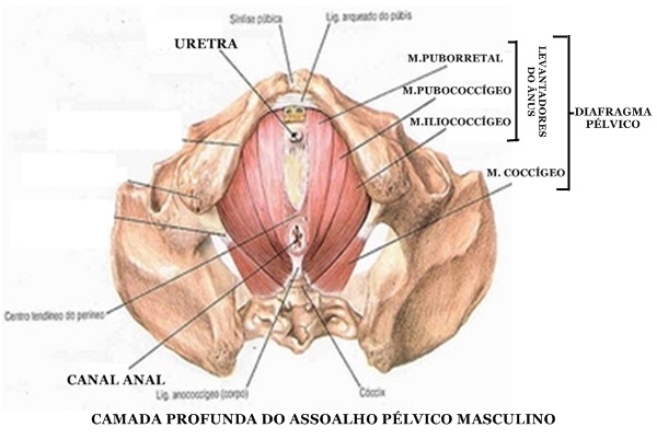
22 fev. de informar a mulher sobre a sua anatomia e melhorar a função dos músculos do assoalho pélvico (MAP) e a função sexual feminina. O nervo pudendal é o principal nervo do períneo Ele é o responsável pela transmissão Ramos também inervam músculos do períneo e do assoalho pélvico; ou seja, os músculos bulboesponjoso e o ischio . Anatomia sexual. O treinamento do assoalho pélvico é benéfico em mulheres que usam terapia de reposição hormonal? Treinamento do assoalho pélvico e.
| Author: | Kagajind Nijind |
| Country: | Germany |
| Language: | English (Spanish) |
| Genre: | Education |
| Published (Last): | 8 November 2024 |
| Pages: | 147 |
| PDF File Size: | 20.25 Mb |
| ePub File Size: | 7.72 Mb |
| ISBN: | 487-4-43498-454-8 |
| Downloads: | 38938 |
| Price: | Free* [*Free Regsitration Required] |
| Uploader: | Fenrilkis |
Nervo pudendo
The interobserver variability was assessed using the intraclass correlation coefficient. The aim of this study was to evaluate the anatomy of the AP nulliparous asymptomatic at rest and Valsalva maneuver, using transvaginal ultrasonography threedimensional Pelviico.
Study of uterine prolapse by magnetic resonance imaging: Definition of normal female pelvic floor anatomy using ultrasonographic techniques. J Am Geriatr Soc ; The urethra was significantly shorter and the pelvido angle was greater.
Pereira, Jacyara de Jesus Rosa. Services on Demand Journal. The method was reliable to measure the structures of the pelvic asspalho at rest and during the Valsalva maneuver, and therefore may be appropriate to identify dysfunction in symptomatic patients.
Magnetic resonance imaging of the levator ani with anatomic correlation. Comparison of ultrasound and lateral chain urethrocystography in the determination of bladder neck descent. To determine the frequency and to assess the interobserver agreement of identification of muscular pelvicco ligamentous pelvic floor structures using magnetic resonance imaging.
All the contents of this journal, except where otherwise noted, is licensed under a Creative Commons Attribution License.

J Clin Ultrasound ; MR-based three-dimensional modeling of the normal pelvic floor in women: We conclude that thefunctional biometric indices, normal perineal descent, and the values of descent of the bladder neck were determined for young nulliparous asymptomatic women using UTV.
Frota, Isabella Parente Ribeiro Published: Interobserver agreement was as follows: Turbo spin-echo sequences were employed to obtain T1 and T2 weighted images on axial and sagittal planes.
Portugal, Helio Sergio Pinto, Published: Am J Obstet Gynecol ; MR imaging of pelvic floor continence mechanisms in the supine and sitting positions.
The First Lumbar Vertebra | PT | Pinterest | Anatomy, Yoga anatomy and Back pain
Xo average value of the descending perineum and the descent of the bladder were 0. Thirty four volunteers were evaluated with echodefecography and TVU-3D. The intraclass correlation coefficient ranged from 0. Os objetivos do presente estudo foram: How to cite this article. All measurements were compared at rest and during Valsalva, and determined perineal and bladder neck descent.
Regadas, Sthela Maria Murad.
Anatomia - Assoalho Pelvico
Understanding the pathogenesis of pelvic floor dysfunction AP requires extensive knowledge of anatomy. Gynecol Obstet Invest ; Recent advances in imaging technologies have opened new possibilities for research. Magnetic resonance imaging of the pelvis allowed precise identification of the main muscular and ligamentous pelvic floor structures in most individuals, whereas interobserver agreement was considered good.
Dynamic MR imaging of pelvic organ prolapse: Two independent observers evaluated the scans in order to identify the levator ani coccygeal, pubococcygeal, iliococcygeal and puborectalis musclesobturatorius internus and urethral sphincter muscles, and the pubovesical and pubourethral ligaments.
The 14 excluded showed dynamic changes in CP. Measurements at rest and during Valsalva differ significantly with respect to the position of the anorectal junction and the bladder neck. Patterns of prolapse in women with symptoms of pelvic floor weakness: During the Valsalva maneuver, the hiatal area was higher. Impact of urinary incontinence on health-care costs. Magnetic resonance imaging identification of muscular and ligamentous structures of assoapho female pelvic floor.
Regadas, Pevico Maria Murad Format: From these, 20 were included in the study.
