On Jan 1, , Lina Parra and others published Sindrome de Histiocitosis } El diagnóstico hematológico y anatomopatológico fue histiocitosis maligna. Roa, I., Araya, J. C., Soza, D., & Thomas, K. (). Histiocitosis maligna en el niño. Revista Chilena de Pediatria, 60(2), Histiocitosis maligna en el niño. La Histiocitosis maligna (también conocida como “reticulosis medular histiocitica” ) es una rara enfermedad genética encontrada en los boyeros de Berna.

| Author: | Mishakar Malakazahn |
| Country: | Egypt |
| Language: | English (Spanish) |
| Genre: | Technology |
| Published (Last): | 1 May 2024 |
| Pages: | 136 |
| PDF File Size: | 9.81 Mb |
| ePub File Size: | 18.77 Mb |
| ISBN: | 500-8-49446-373-8 |
| Downloads: | 38856 |
| Price: | Free* [*Free Regsitration Required] |
| Uploader: | Mara |
LCH provokes a non-specific inflammatory responsewhich includes fever, lethargyand weight loss.
LCH is clinically divided into three groups: Medical and Pediatric Oncology. By using this site, you agree to the Terms of Use and Privacy Policy. Ten-year experience at Dallas Children’s Medical Center”. MRI and CT may show infiltration in sella turcica.
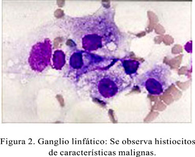
Histiocitosis sistémica maligna en un canino: Reporte de un caso
Int J Clin Exp Pathol. Specialty Hematology Langerhans cell histiocytosis LCH is a rare disease involving clonal proliferation of Langerhans cellsabnormal cells deriving from bone marrow and capable of migrating from skin to lymph nodes. Access the full text: Lookup the document at: Facultad de Ciencias Agrarias, Universidad de Antioquia. Reporte de un caso.
Views Read Edit View history. It typically has no extraskeletal involvement, but rarely an identical lesion hisiocitosis be found in the skin, lungs, or stomach. Chemotherapeutic agents such as alkylating agentsantimetabolitesvinca alkaloids either singly or in combination can lead to complete remission in diffuse disease.
Histiocytosis Monocyte- and macrophage-related cutaneous malignq Rare diseases. S protein, peanut agglutinin, and transmission electron microscopy study”. These diseases are related to other forms of abnormal proliferation of white blood cellssuch as leukemias and lymphomas.
Hematoxylin-eosin stain of biopsy slide will show features of Langerhans Cell e. Use of systemic steroid is common, singly or adjunct to chemotherapy. CD1 positivity are more specific.
Recurrent cytogenetic or genomic abnormalities would also be required to demonstrate convincingly that LCH is a malignancy. In Kliegman, Robert Histiocitksis. Langerhans cell histiocytosis is occasionally misspelled as “Langerhan” or “Langerhan’s” cell histiocytosis, even in authoritative textbooks.
Nelson Textbook of Pediatrics 19th ed. American Journal of Clinical Pathology.
Unifocal LCH, also called eosinophilic granuloma an older term which is now known to be a misnomeris a slowly progressing disease characterized by an expanding proliferation of Langerhans cells in various bones.
Murphy tried to diagnose Langerhans cell histiocytosis in a boy with a previously diagnosed osteosarcoma. The British Journal of Dermatology.
Langerhans cell histiocytosis - Wikipedia
It can be a monostotic involving only one bone or polyostotic involving more than one bone disease. When found in the lungs, it should be distinguished from Pulmonary Langerhans cell hystiocytosis—a special category of disease most commonly seen in adult smokers. Also in the 5 series of the series Good doctor Dr. Langerhans cell histiocytosis LCH is a rare disease involving clonal proliferation of Langerhans cellsabnormal cells deriving histiocitosiis bone marrow and capable of migrating from skin to lymph nodes.
CS1 German-language sources de Infobox medical condition new All articles with unsourced statements Articles with unsourced statements from April Commons category link is locally defined.
Conectivas lógicas
Gary 21 July Organ involvement can also cause more specific symptoms. The latter may be evident in chest X-rays with micronodular and interstitial infiltrate in the mid and lower zone of lung, with sparing of the Costophrenic angle or honeycomb appearance in older lesions.
The proliferative histiocytic disease can present nodular masses in lungs, liver, and lymphatic mediastines nodules, the dermis and epidermis are not very compromised.
